Not directly. They are different manifestations of venous disease but share common underlying risk factors. Someone with spider veins may be at higher risk for varicose veins, but one does not transform into the other.
Symptom Guide
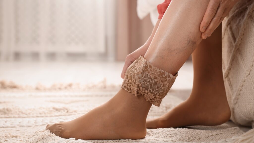
Telangiectasias on Legs: Causes, Symptoms, Diagnosis & Treatment
Telangiectasias on legs (spider veins): what causes them, how they’re diagnosed, and the most effective treatments. Learn when to see a vein specialist.


Key takeaways
- Telangiectasias (spider veins) are dilated intradermal venules less than 1 mm in diameter, classified as C1 in the CEAP system for chronic venous disorders.
- They are extremely common, affecting 79–88% of adults.
- Risk factors include family history, female sex, pregnancy, hormonal medications, prolonged standing or sitting, obesity, and aging.
- Telangiectasias can be an early visible sign of chronic venous insufficiency (CVI), particularly when accompanied by symptoms such as leg heaviness, aching, or swelling.
- Sclerotherapy is the first-line treatment, while laser vein therapy is an effective alternative for very small vessels.
- Duplex ultrasound is recommended before treatment to rule out underlying venous reflux.
Table Of Contents
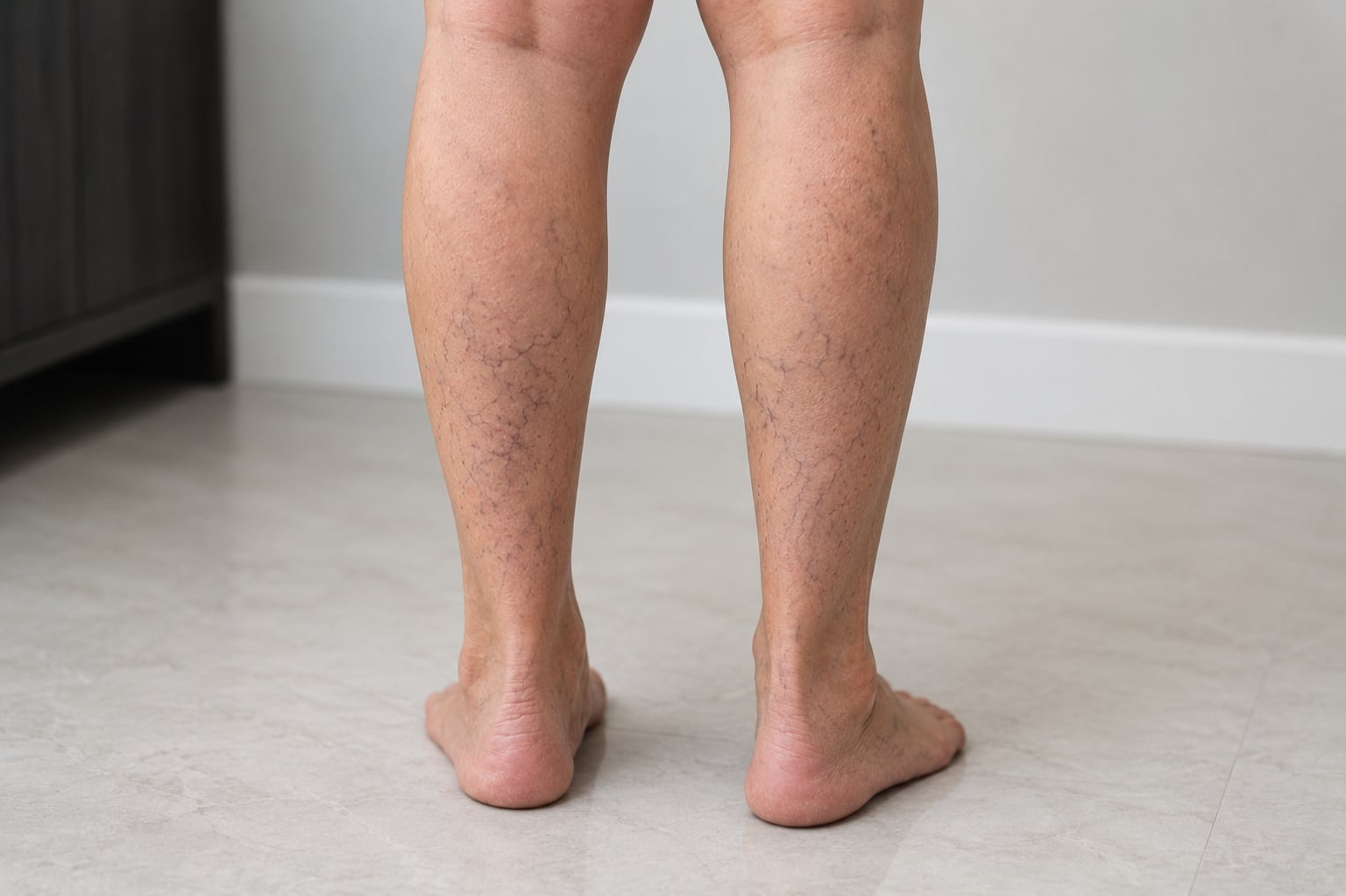
Telangiectasias — commonly called spider veins — are small, dilated blood vessels less than 1 mm in diameter that appear as thin red, blue, or purple lines just beneath the skin’s surface. They most often develop on the legs and are among the most common vascular findings in adults.
A population study of over 1,300 adults found that 88% of women and 79% of men had visible telangiectasias on their legs.
Although spider veins are frequently regarded as a cosmetic concern, they may also indicate underlying venous disorders that require medical assessment. Symptoms such as aching, burning, itching, or leg heaviness can occur and extend beyond cosmetic considerations. This resource outlines the definition, causes, diagnostic methods, and available treatments for telangiectasias.
What are telangiectasias on legs?
Telangiectasias are permanently dilated small blood vessels — venules, capillaries, or arterioles — visible through the skin. The term was coined by von Graf in 1807, from the Greek words telos (end), angeion (vessel), and ektasis (dilation). Other common names include spider veins, thread veins, sunburst veins, and venous flares.
Red telangiectasias tend to be more superficial and contain oxygenated blood, while blue or purple ones sit slightly deeper and carry deoxygenated blood. The most common locations are the posteromedial thigh, behind the knee (popliteal fossa), and the upper calf.
Spider veins vs. Telangiectasias
These terms describe the same condition. “Spider veins” is the informal name used in consumer health resources. “Telangiectasias” is the clinical term used in medical literature and by healthcare providers.
Common patterns on the legs
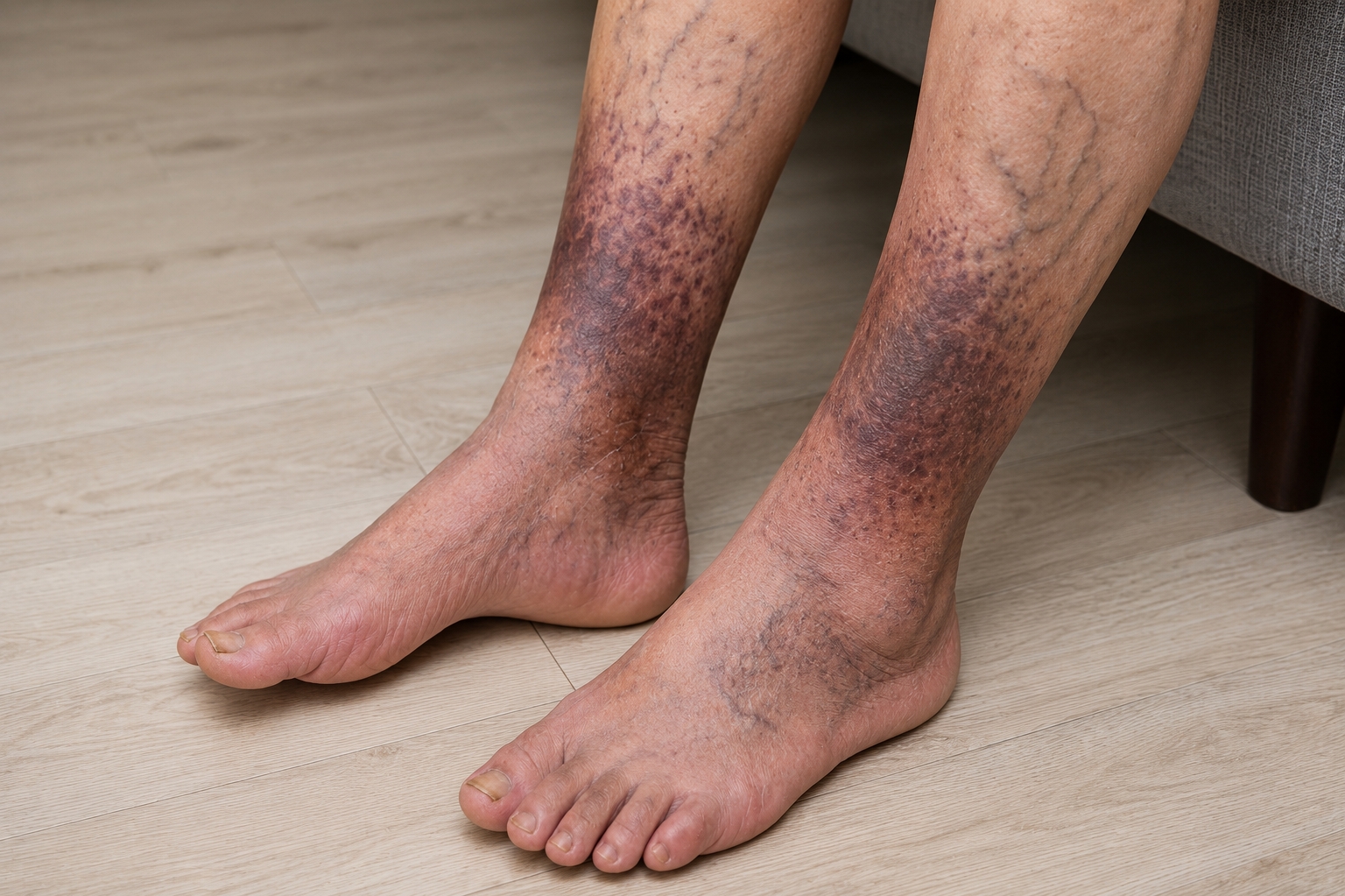
Telangiectasias follow predictable distribution patterns. The Edinburgh Vein Study identified the posteromedial thigh, the popliteal fossa, and the upper calf as the most frequently affected areas. When they appear in a fan-shaped pattern around the ankle — called corona phlebectatica or ankle flare — it may indicate more advanced venous disease. The 2020 CEAP update reclassified corona phlebectatica as C4c, recognizing it as a marker of chronic venous insufficiency.
Prevalence
Telangiectasias are among the most common vascular findings in adults. The most common age for initial presentation is between 30 and 50 years, though they can appear at any age. Women are affected slightly more often than men, largely due to hormonal influences.
Causes and risk factors
The pathophysiology of telangiectasias is not fully understood. A 2018 systematic review concluded that their pathophysiology and risk factors remain an area of active research. Several well-established contributing factors have been identified.
Underlying venous reflux and feeder veins
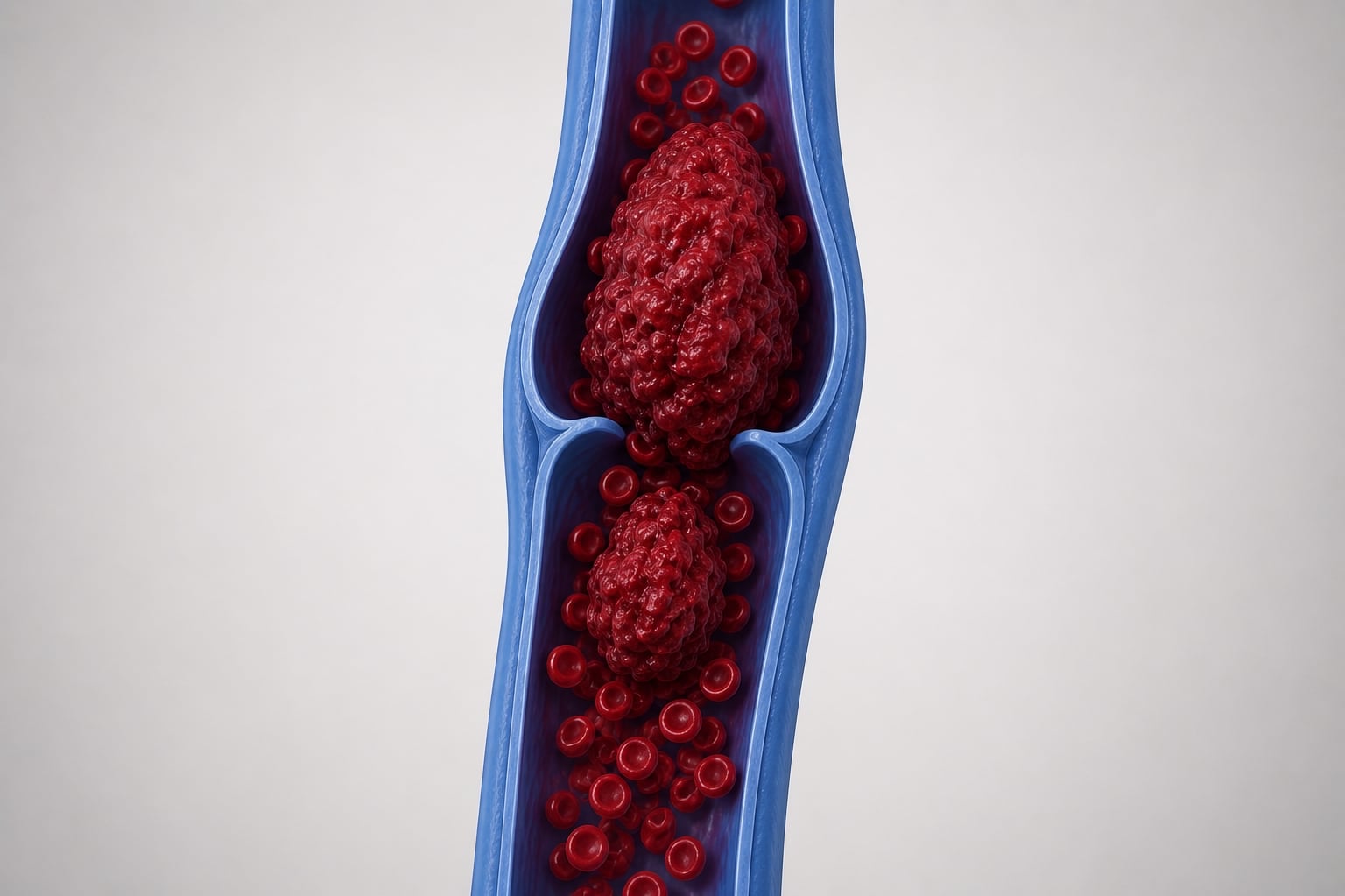
There is a consistent association between spider veins and underlying venous reflux. A sonographic study of thigh telangiectasias found that all 43 affected limbs had incompetent reticular veins underlying the spider vein sites. Reticular veins (1–3 mm) sit just below the skin and act as “feeder” veins — when their valves malfunction, blood flows backward, creating pressure that dilates the surface vessels.
This connection explains why treating telangiectasias without addressing the underlying feeder veins often leads to recurrence.
Genetics, hormones, and age
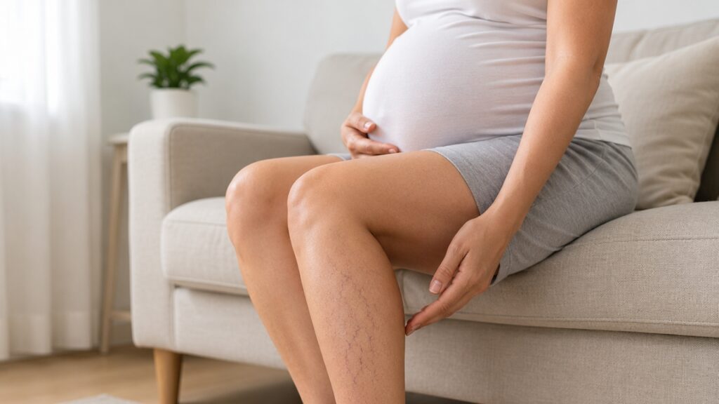
Family history plays a significant role. Hereditary variations in vein wall structure and valve function increase susceptibility. Estrogen and progesterone weaken vein walls and valves, which is one reason telangiectasias are more prevalent in women. Pregnancy creates a combined effect: hormonal changes relax the vein walls while the growing uterus increases pressure on pelvic veins. Hormonal contraceptives and hormone replacement therapy can have similar effects. Aging naturally weakens the integrity of valve and vein walls.
Lifestyle factors
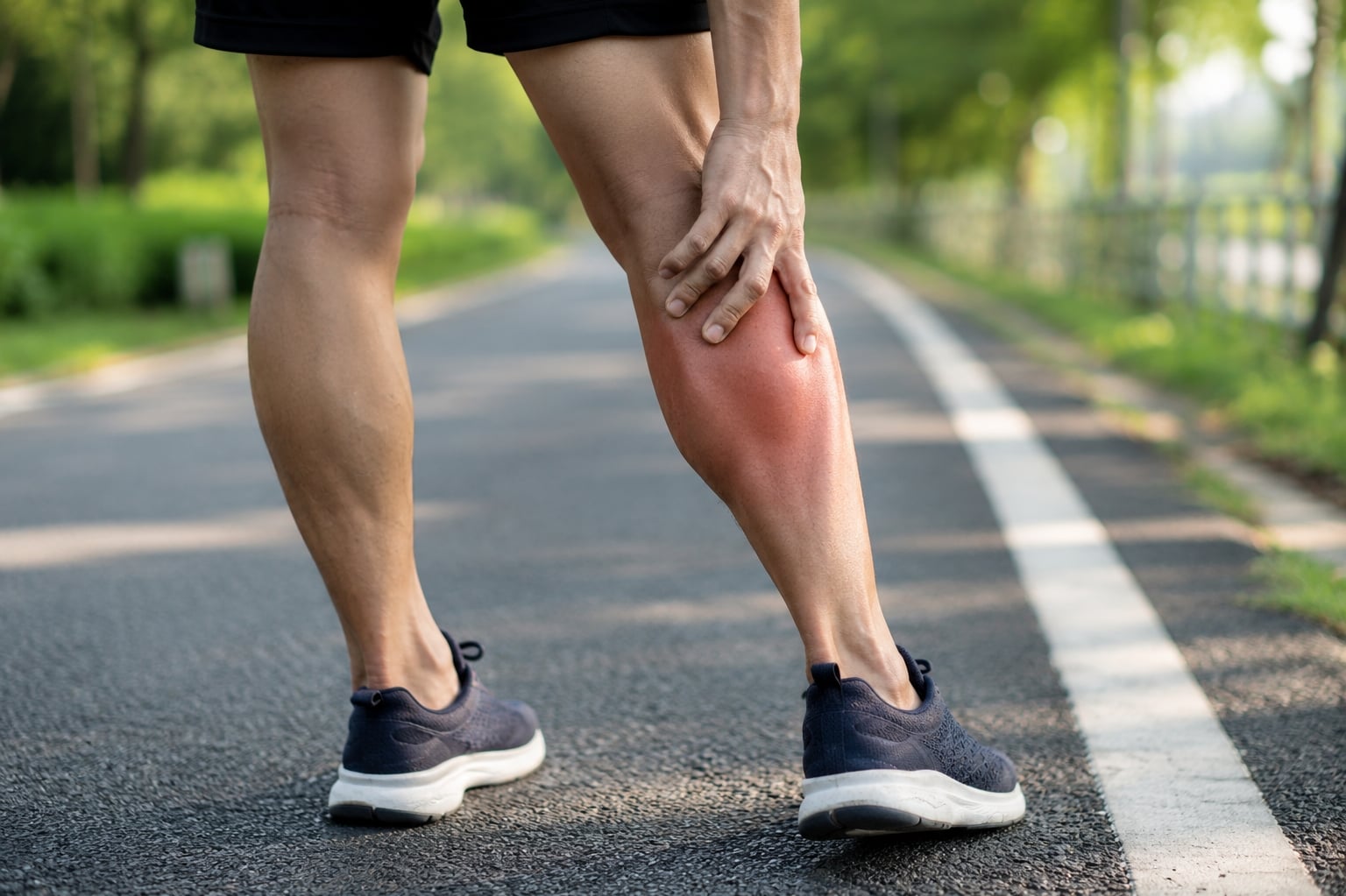
Prolonged standing or sitting reduces the efficiency of the calf muscle pump — the mechanism that pushes blood upward against gravity. Occupations that require prolonged standing or desk work are associated with a higher risk of venous disease. Obesity places additional pressure on leg veins and is linked to a higher prevalence of incompetent perforator veins underlying telangiectasias.
Other contributing factors
Chronic sun exposure can damage superficial blood vessels. Direct trauma, prolonged heat exposure (hot tubs, saunas), and certain medications — particularly long-term topical corticosteroids — can also contribute. In rare cases, telangiectasias on the legs may be associated with systemic conditions such as liver disease, connective tissue disorders, or hereditary hemorrhagic telangiectasia (HHT). For most people, however, leg telangiectasias are associated with venous insufficiency and the risk factors above.
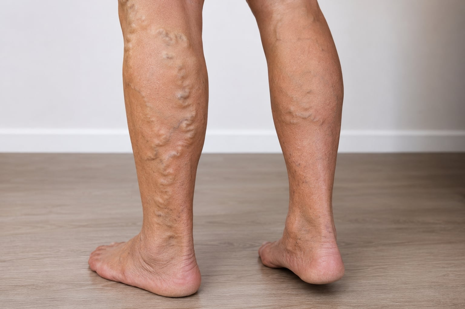
Telangiectasias vs. Reticular veins vs. Varicose veins
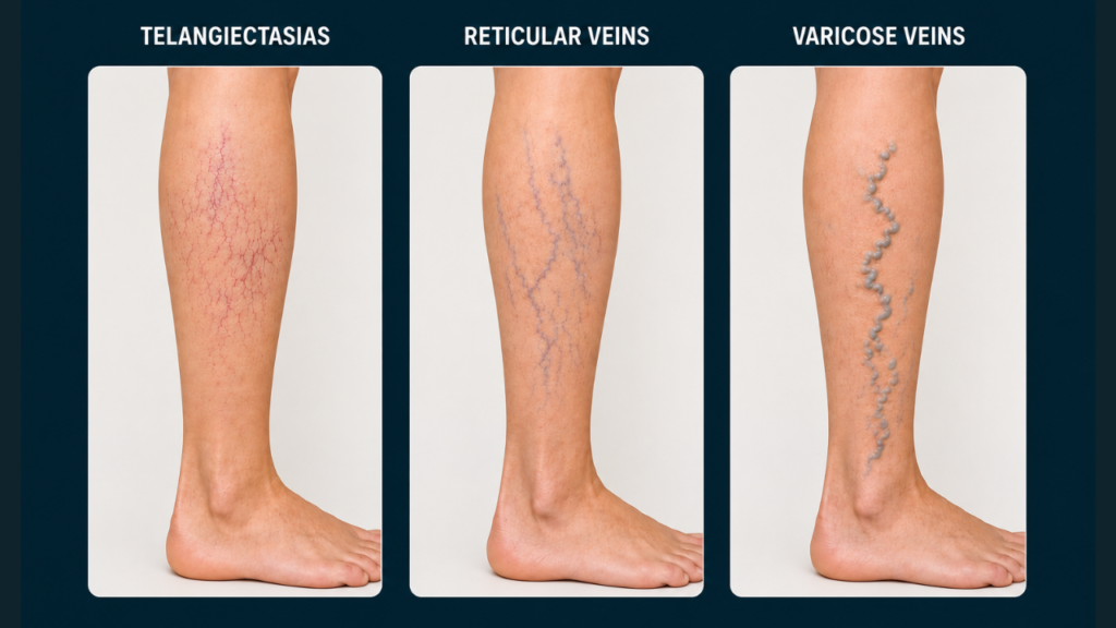
Telangiectasias, reticular veins, and varicose veins differ in size, depth, appearance, and treatment options. Understanding these differences can help identify the most suitable treatment for each condition:
- Telangiectasias (Spider veins): These are very small veins, typically measuring less than 1 mm in diameter. They are located within the skin (intradermal) and usually appear as thin, web-like clusters. Their color can be red, blue, or purple. Classified as C1 under the CEAP system, telangiectasias are commonly treated with sclerotherapy or laser vein therapy.
- Reticular veins: These veins are slightly larger than telangiectasias, measuring between 1–3 mm in diameter. They are found just beneath the skin (subdermal) and often appear as thread-like veins in blue or green. Like spider veins, reticular veins are classified as C1 and are most frequently treated with sclerotherapy.
- Varicose veins: These veins are larger, with a diameter greater than 3 mm, and are located deeper under the skin (subcutaneous). They often appear bulging, twisted, or rope-like and can be blue, purple, or flesh-colored. Classified as C2, varicose veins typically require more advanced treatments, such as radiofrequency ablation, VenaSeal, or surgical procedures.
Corona phlebectatica (Ankle flare)
Corona phlebectatica is a fan-shaped pattern of blue telangiectasias around the inner or outer ankle. Unlike ordinary spider veins elsewhere on the leg, it is considered an important marker of chronic venous disease. The 2020 CEAP update reclassified it as C4c, reflecting consensus that ankle flare indicates significant underlying venous hypertension and warrants specialist evaluation.
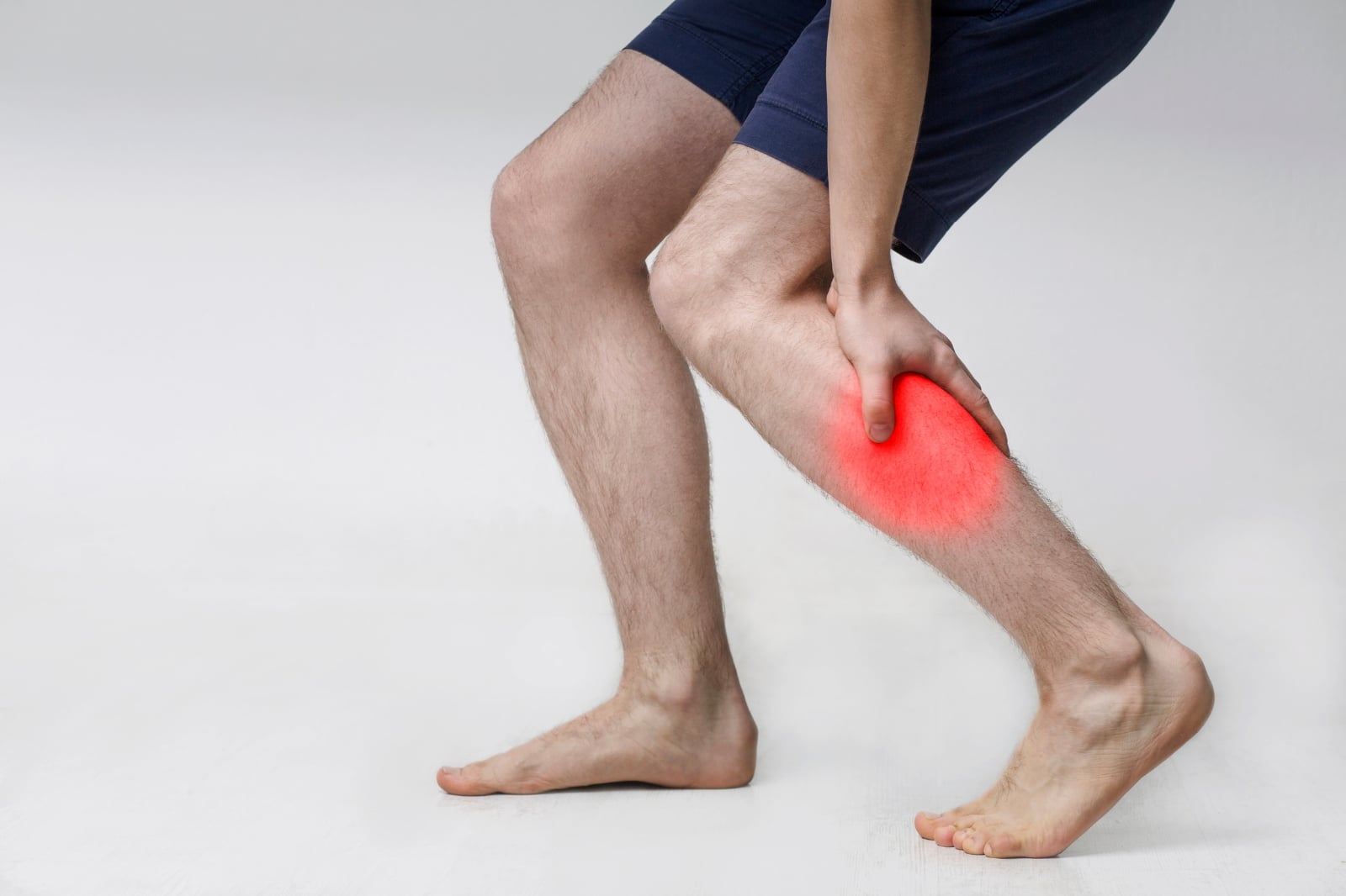
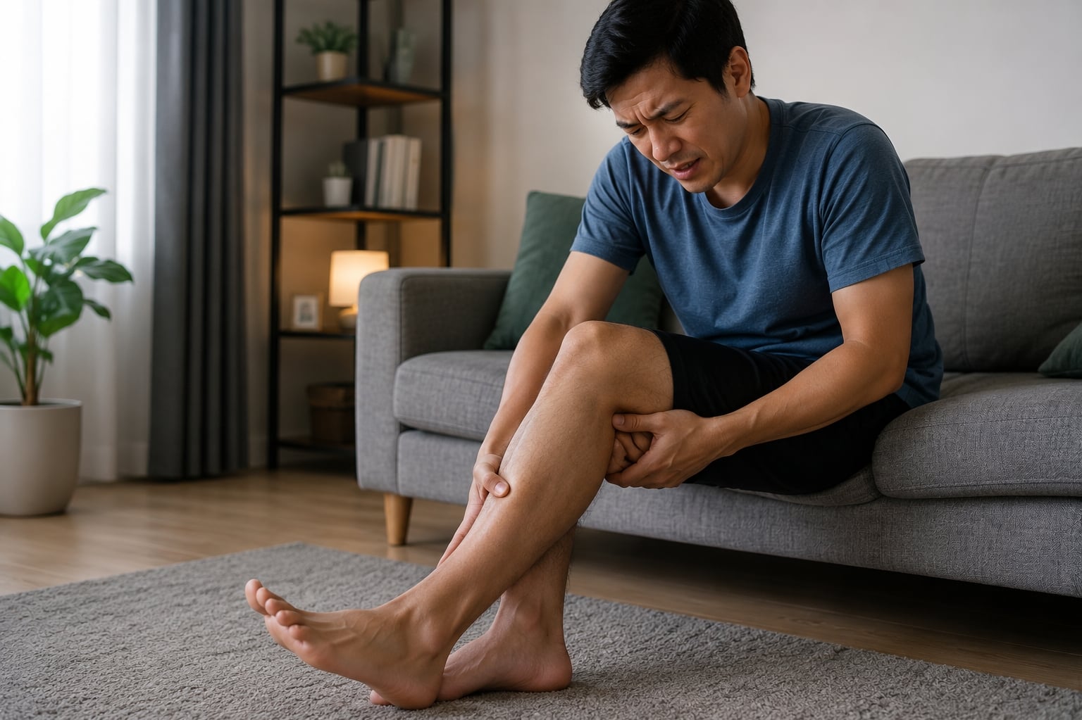
Symptoms and possible complications

Cosmetic concerns vs. Physical discomfort
Many people seek evaluation for cosmetic reasons. However, symptoms can include dull aching, burning, itching, leg heaviness (especially late in the day or after prolonged standing), and nighttime cramps. Symptoms typically worsen with prolonged standing or sitting and improve with leg elevation or walking.
Potential complications
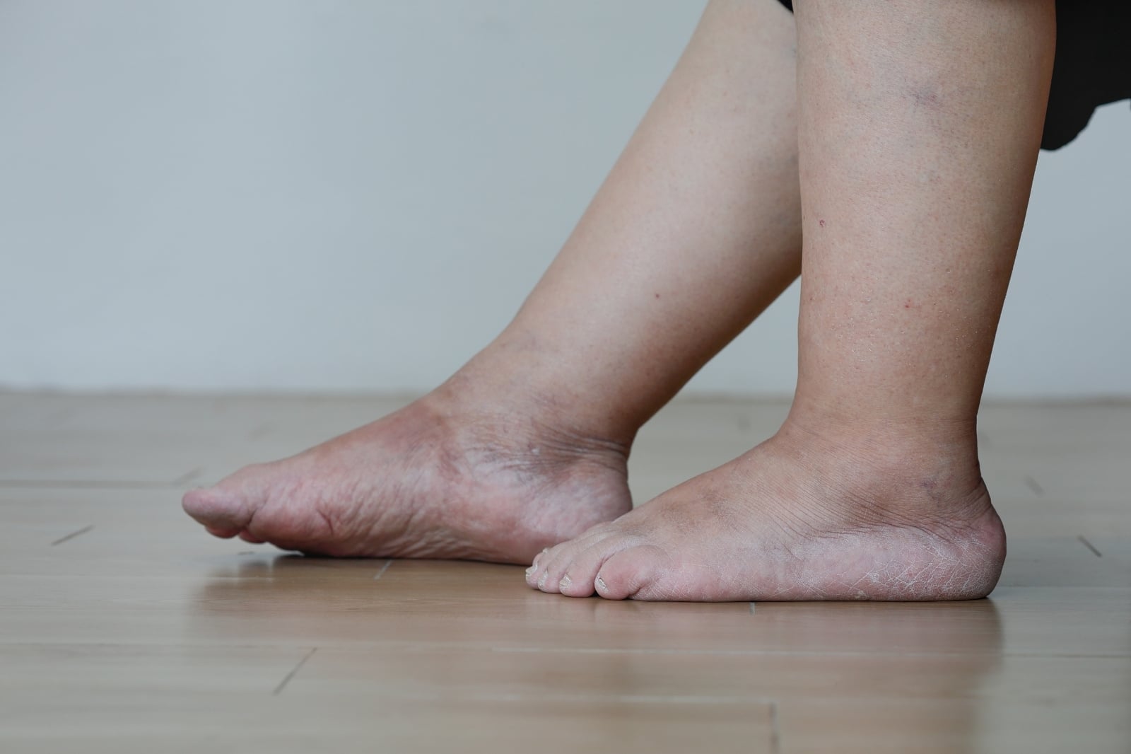
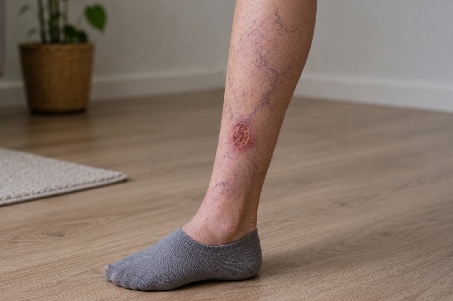
When telangiectasias coexist with chronic venous insufficiency, additional complications can develop: hyperpigmentation around the ankles (from iron deposits in leaked red blood cells), telangiectatic matting (new clusters of fine spider veins), minor bleeding from superficial veins, and — in advanced, untreated venous disease — skin breakdown leading to venous ulcers.
When to see a doctor or vein specialist
Certain signs warrant prompt evaluation:
- Sudden leg swelling
- Warmth, redness, or tenderness along a vein
- Skin darkening around the ankles
- Hard, thickened, or leathery skin on the lower legs
- A wound or sore that does not heal
- Bleeding from a vein
- Fan-shaped vein clusters around the ankles (corona phlebectatica
These symptoms may indicate more advanced venous disease or deep vein thrombosis (DVT).
Pregnancy-related telangiectasias
Spider veins frequently develop or worsen during pregnancy. In most cases, they improve within three to six months following delivery. Vein specialists typically advise waiting at least three to six months postpartum before initiating treatment.
How telangiectasias on legs are diagnosed
History and physical exam
A vein specialist evaluates the patient’s medical history, including family history of venous disease, pregnancies, hormone use, occupation, and symptoms. The physical examination is optimally conducted with the patient standing, as this position accentuates venous filling and reveals abnormalities more clearly.
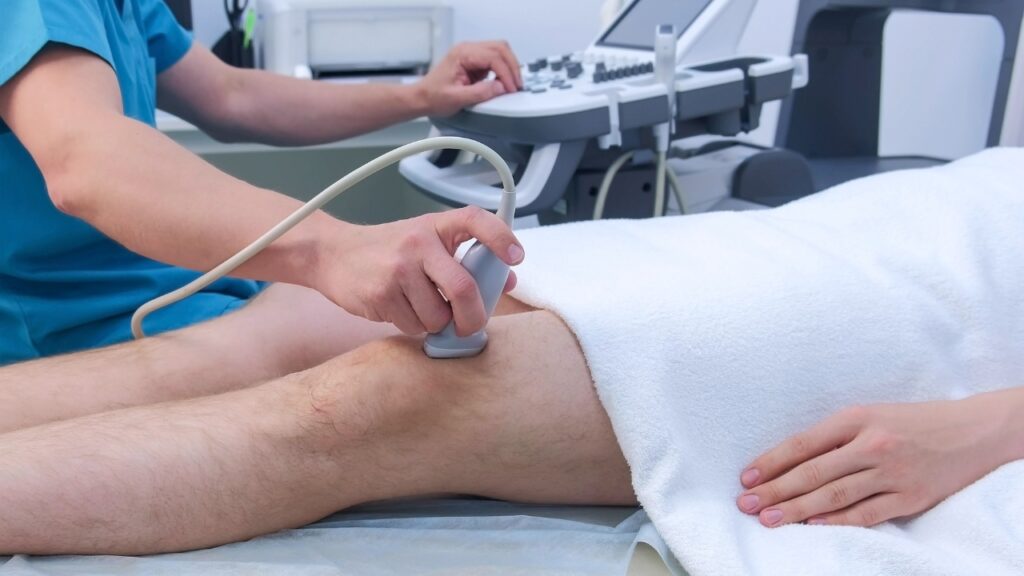
Duplex ultrasound
Duplex ultrasound is the definitive diagnostic tool. This painless, noninvasive test combines ultrasound imaging with Doppler technology to assess blood flow direction and speed. It can identify valve dysfunction (reflux), locate incompetent feeder veins, and rule out DVT. Experts recommend duplex ultrasound before treating leg telangiectasias to improve treatment outcomes and reduce recurrence.
CEAP classification
The CEAP classification system (Clinical, Etiological, Anatomical, Pathophysiological) standardizes the description of chronic venous disorders. Telangiectasias and reticular veins are classified as C1. Each class is designated as symptomatic (S) or asymptomatic (A) — for example, C1S indicates spider veins with aching or itching.
Differential diagnosis
Conditions that can mimic telangiectasias include cherry angiomas, petechiae or purpura (which do not blanch with pressure), capillary malformations (port-wine stains), and hemangiomas.
Proven treatments for leg telangiectasias
Conservative measures
Graduated compression stockings enhance venous return and decrease blood pooling. Regular physical activity, such as walking, cycling, and calf raises, stimulates the calf muscle pump. Elevating the legs above heart level for 15 to 20 minutes multiple times daily can alleviate discomfort.
Sclerotherapy (First-line)
Sclerotherapy is the gold standard treatment. A Cochrane systematic review confirmed its superiority over placebo. A sclerosing solution — most commonly polidocanol or sodium tetradecyl sulfate (STS) — is injected into the affected veins. The solution causes the vein walls to collapse, and the body gradually absorbs the treated vein over several weeks.
Liquid sclerotherapy is the standard treatment for telangiectasias less than 1 mm in diameter. Foam sclerotherapy, which involves mixing the sclerosant with gas to form a microfoam, is more effective for feeder reticular veins measuring 1 to 3 mm. Utilizing both techniques in combination yields superior outcomes compared to either method alone.
Most patients require two to four treatment sessions, scheduled at intervals of four to six weeks. The Mayo Clinic reports closure rates of 80% to 90%, with treated veins generally disappearing within six to eight weeks.
Temporary hyperpigmentation develops in 10% to 30% of patients and typically resolves within six to twelve months. Less common adverse effects include telangiectatic matting, minor skin ulceration at the injection site, allergic reactions, and deep vein thrombosis, which occurs in fewer than 1% of cases.
Laser vein therapy
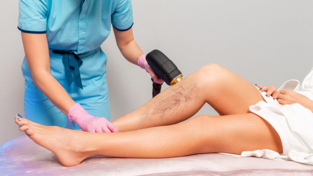
Laser vein therapy is the most widely studied for leg telangiectasias. A comparative study of 285 women found that the laser vein removal was most effective for telangiectasias measuring less than 1 mm, while sclerotherapy with polidocanol was more effective for larger vessels. The 1064 nm wavelength has low melanin absorption, making it suitable for a range of skin types, including darker skin tones.
Pulsed dye lasers (PDL) and intense pulsed light (IPL) are generally less effective on leg veins than on facial telangiectasias due to the greater depth and higher pressure of leg vessels.
Thermocoagulation and radiofrequency ablation
Ohmic thermolysis delivers focused heat via a fine-needle probe. It is best suited for very fine, superficial telangiectasias that are too small to be injected or too superficial for laser.
Treating feeder veins
Because telangiectasias are frequently connected to incompetent reticular and perforator veins, treating only the visible surface vessels often leads to recurrence. Options include microphlebectomy (removal of reticular veins through small skin punctures) and endovenous ablation (radiofrequency or laser energy to close incompetent saphenous veins).
Combination therapy
Evidence supports a combined approach. Low-certainty evidence from a 2021 Cochrane Review suggests that laser plus sclerotherapy may produce more resolution than sclerotherapy alone. A typical treatment sequence: correct saphenous reflux with endovenous ablation first, treat feeder reticular veins with foam sclerotherapy, then address residual telangiectasias with liquid sclerotherapy or laser vein therapy.
Recovery and aftercare
Following sclerotherapy, graduated compression stockings are generally worn for one to three weeks. Ambulation is encouraged immediately after the procedure. High-impact exercise, heavy lifting, and prolonged standing should be avoided for several days. Hot baths, saunas, and direct sun exposure should be avoided for at least two weeks to reduce the risk of hyperpigmentation. If skin darkening occurs, it typically fades gradually over several months.
Prevention and self-care
- Take movement breaks every 30 minutes during prolonged sitting or standing.
- Incorporate calf raises and walking into daily routines.
- Maintain a healthy weight.
- Elevate the legs when resting.
- Wear graduated compression stockings during long flights, road trips, or standing-intensive work.
- Apply broad-spectrum sunscreen to exposed legs and avoid excessive heat exposure.
Special situations
- Pregnancy and breastfeeding: Sclerotherapy and laser treatments are not performed during either. Treatment is generally deferred until three to six months postpartum.
- Skin color: Darker skin tones carry a higher risk of post-inflammatory hyperpigmentation after sclerotherapy or laser therapy. The laser vein therapy is the safest choice due to its low melanin absorption. Conservative energy settings and test spots are recommended.
- Athletes: Low-impact exercise can typically resume within 24–48 hours after sclerotherapy. High-intensity or competitive activities should be avoided for five to seven days.
- Blood thinners, diabetes, and PAD: These conditions may affect treatment planning and compression therapy. Treatments can generally proceed with appropriate modifications.
Costs, insurance, and expectations
When spider veins are purely cosmetic (no symptoms, no underlying disease), treatment is typically not covered by insurance. When telangiectasias are symptomatic or associated with documented venous insufficiency on duplex ultrasound, some or all treatment costs may be covered. Sclerotherapy typically ranges from $300–$600 per session; laser treatment ranges from $400–$900 per session. Most patients need two to four sessions.
Frequently asked questions
Treated veins do not reopen. However, new telangiectasias may develop over time, especially if underlying risk factors persist. Addressing feeder veins and venous reflux significantly reduces recurrence.
In the vast majority of cases, no. They are related to venous insufficiency, genetics, and lifestyle factors. In rare instances, they can be associated with connective tissue disorders such as scleroderma or lupus.
No topical product or supplement has been scientifically proven to close or remove dilated vessels. Sclerotherapy and laser remain the only evidence-based methods.
They can be. The Edinburgh Vein Study found a highly significant association between the severity of telangiectasias and the severity of varicose veins. Duplex ultrasound can determine whether they reflect underlying CVI.
Sclerotherapy involves a mild pinching or stinging sensation. Each session takes 15–30 minutes. The laser vein removal produces a snapping or warming sensation; topical numbing cream can be applied. Both treatments are well-tolerated, and normal activities can resume the same day.
Bottom line
Telangiectasias on the legs are extremely common and usually harmless, but they can indicate underlying chronic venous insufficiency. A duplex ultrasound evaluation is the most important step in determining whether deeper venous disease is contributing to the problem.
Sclerotherapy remains the proven first-line treatment, and combination approaches — addressing feeder veins and underlying reflux before treating the visible spider veins — produce the most lasting results. With proper evaluation and targeted treatment, most people achieve significant improvement in both appearance and comfort.
A consultation with a vein specialist, such as those at iThriveVeins, is recommended for comprehensive vein evaluation and individualized treatment planning.
- ScienceDirect. 2008. Telangiectasia in the Edinburgh Vein Study.
- National Institutes of Health. 2024. Spider Veins.
- Journal of Vascular Surgery. 2020. The 2020 update of the CEAP classification system and reporting standards.
- National Institutes of Health. 2024. Treatment for telangiectasias and reticular veins.
- National Institutes of Health. 2018. Pathophysiology of telangiectasias of the lower legs.
- National Institutes of Health. 2018. A sonographic study of thigh telangiectasias.
- National Institutes of Health. 2023. CEAP Classification of Venous Disorders.
- PubMed. 2011. Sclerotherapy for lower limb telangiectasias.
- MayoClinic. 2011. Percutaneous Treatment of Varicose Veins Offers Less Invasive, More Effective Options.
- PubMed Central. 2018. Comparative study in leg telangiectasias treatment with laser and sclerotherapy.
- PubMed. 2021. Treatment for telangiectasias and reticular veins.
Editorial standards
All iThriveVeins content is medically reviewed by board-certified vein specialists and written following evidence-based guidelines. We source our information from peer-reviewed medical journals, clinical studies, and established medical organizations. Our editorial process ensures accuracy, objectivity, and relevance to patient needs.