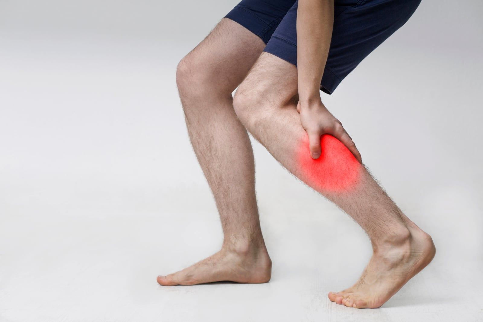
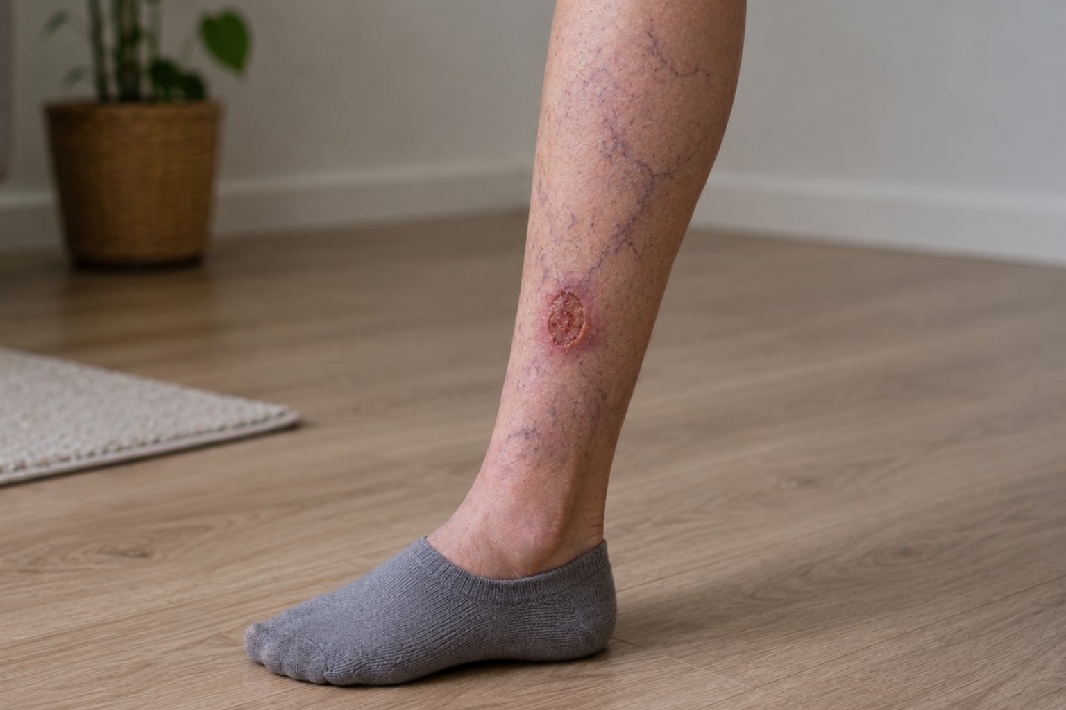
A venous ulcer is typically a shallow, irregularly shaped wound on the inner lower leg or ankle. The wound bed is usually pink or yellowish with moderate drainage. The surrounding skin often shows brown discoloration, hardening, or eczema-like changes.
Symptom Guide
Open Wounds on Legs: Venous Ulcers, Causes & Treatment
Open leg wounds often result from venous disease. This article discusses venous ulcer development, factors contributing to impaired healing, & treatment options.


Key takeaways
- Venous leg ulcers represent the most prevalent form of chronic leg wound, accounting for 60–80% of all non-healing leg ulcerations.
- These ulcers result from chronic venous hypertension, defined as sustained elevated pressure in the leg veins, which initiates inflammation, tissue damage, and impaired wound healing.
- Accurate diagnosis is essential because leg wounds can also stem from arterial disease, diabetes, or mixed etiologies.
- Compression therapy serves as the foundation of care to counteract venous hypertension and reduce swelling.
- Early endovenous ablation of superficial venous reflux significantly accelerates healing and lowers recurrence rates.
- Preventing ulcer recurrence requires lifelong compression, definitive venous treatments, daily leg elevation, and regular physical activity.
Table Of Contents
An open wound on the leg that fails to heal over weeks, months, or even years is often a venous leg ulcer (VLU). These chronic wounds arise when prolonged elevated pressure within the leg veins leads to skin and tissue breakdown. Venous ulcers constitute 60–80% of all chronic leg ulcerations and represent the most advanced clinical manifestation of chronic venous disease.
A 2021 systematic review of international data estimated the prevalence of VLUs at approximately 1.69% and the incidence at up to 1.33%. In the United States, an estimated 500,000 individuals have active venous ulcers, resulting in substantial healthcare expenditures. Despite available treatments, only about 22% of venous ulcers heal within three months, and recurrence rates remain elevated, reaching up to 78% within three years in the absence of ongoing preventive care.
What causes open wounds on the legs?
Not all leg wounds are venous ulcers. Open wounds on the legs may result from various etiologies, and accurate identification of the underlying cause is essential for effective management.
Venous disease (Most common)
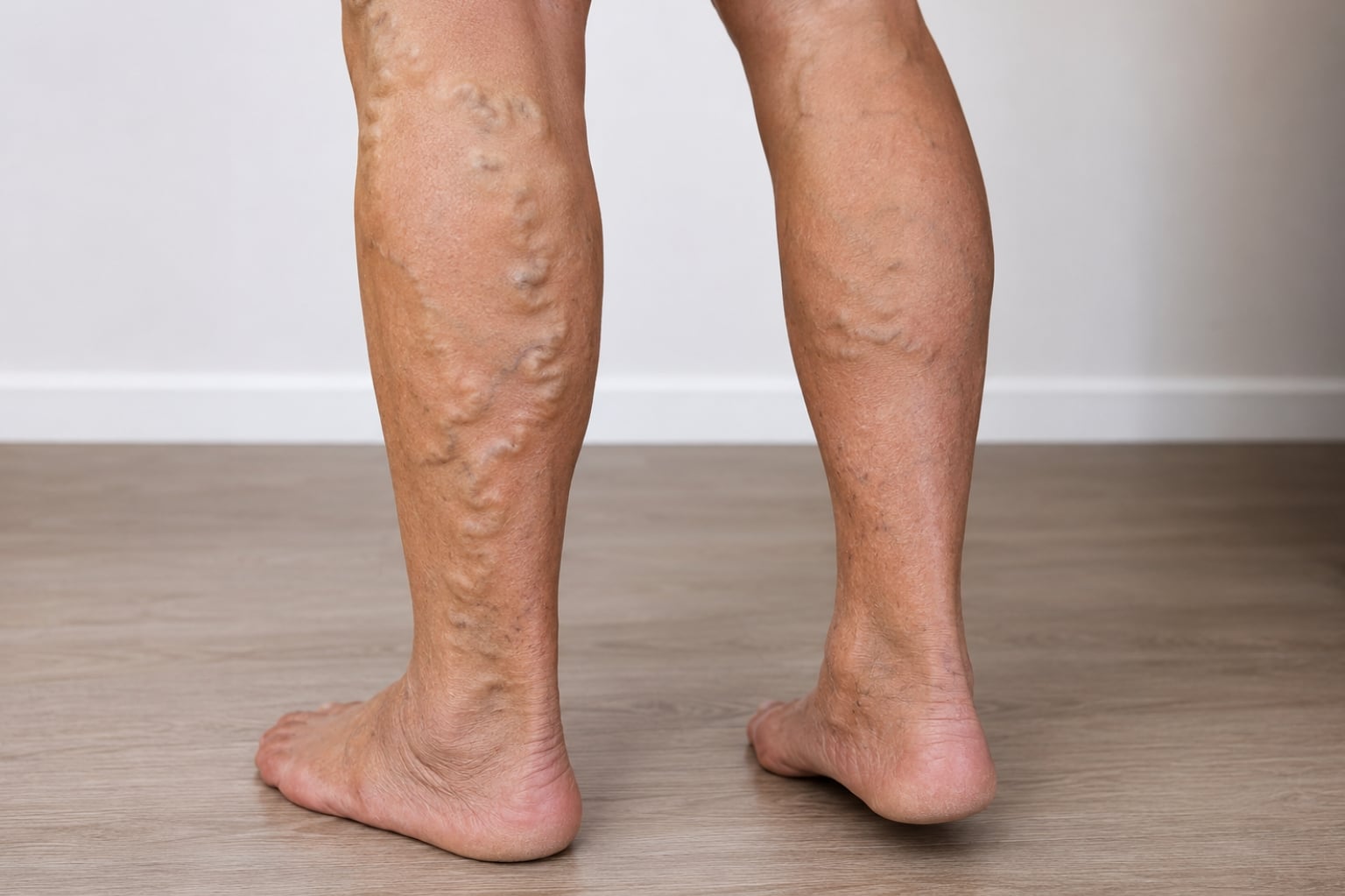
Chronic venous insufficiency (CVI) is the primary cause of non-healing leg wounds. Malfunctioning vein valves result in blood pooling in the lower extremities, leading to sustained venous hypertension. This condition initiates a chronic inflammatory cascade: red blood cells leak into surrounding tissues and release iron, white blood cells infiltrate the dermis and release matrix metalloproteinases, and fibroblasts adopt an abnormal contractile phenotype that increases skin tension rather than facilitating repair. The consequence is tissue breakdown, manifesting as an open, typically shallow wound on the inner ankle or lower leg that resists healing.
Venous ulcers typically present in the gaiter zone, defined as the area between the ankle and mid-calf, most frequently on the medial (inner) aspect of the leg. These ulcers often exhibit irregular borders, a shallow wound bed containing pink or yellow granulation tissue, and surrounding skin with features of chronic venous disease, such as brown discoloration (hemosiderin staining), hardened or leathery texture (lipodermatosclerosis), or eczema-like inflammation (stasis dermatitis).
Arterial disease
Peripheral artery disease (PAD) may result in leg wounds when arterial blood flow is severely compromised. Arterial ulcers commonly develop on the toes, feet, or bony prominences such as the shins and heels. These ulcers are characterized by a punched-out appearance with well-defined edges, a pale or necrotic wound bed, and minimal exudate. The surrounding skin is typically pale, cool, and hairless. Arterial ulcers are often painful, especially at night or when the legs are elevated, in contrast to venous ulcers, which improve with elevation.
Diabetes
Diabetic foot ulcers arise from a combination of peripheral neuropathy (loss of protective sensation), microvascular disease, and impaired immune function. These ulcers most frequently develop on the plantar surface (sole) of the foot at sites of increased pressure.
Mixed etiology
In many patients, particularly older adults, venous disease, arterial disease, and diabetes may coexist. A wound management review emphasized that VLUs can have mixed etiologies, necessitating treatment strategies that address all contributing factors. The ankle-brachial index (ABI) is a critical screening tool for assessing arterial sufficiency prior to initiating compression therapy.
Other causes
Less common etiologies of leg wounds include traumatic injury with impaired healing, inflammatory conditions such as pyoderma gangrenosum and vasculitis, infection, lymphedema, malignancy, and radiation injury. A comprehensive diagnostic evaluation is essential to avoid misattributing a wound to venous disease when alternative causes may require distinct management approaches.
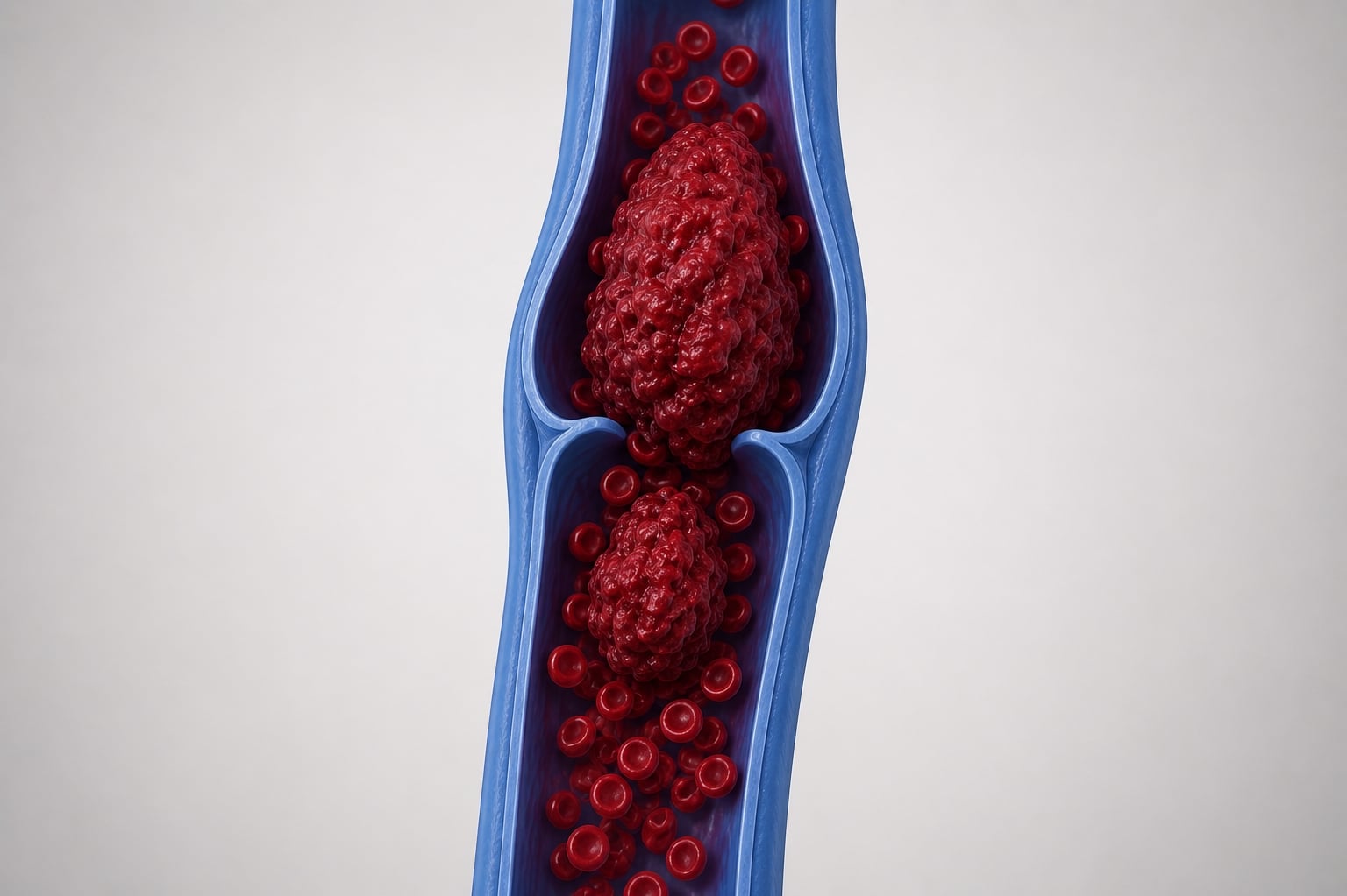
How venous disease leads to open wounds

The pathological cascade
The development of a venous ulcer follows a well-characterized sequence. It begins with venous valve incompetence — valves in the superficial, deep, or perforator veins fail to prevent backflow. Blood refluxes and pools, producing ambulatory venous hypertension. A review of VLU pathophysiology in JVSV describes the downstream events: elevated venous pressure is transmitted to the microcirculation, where it injures capillary endothelium. Capillaries become dilated, tortuous, and abnormally permeable.
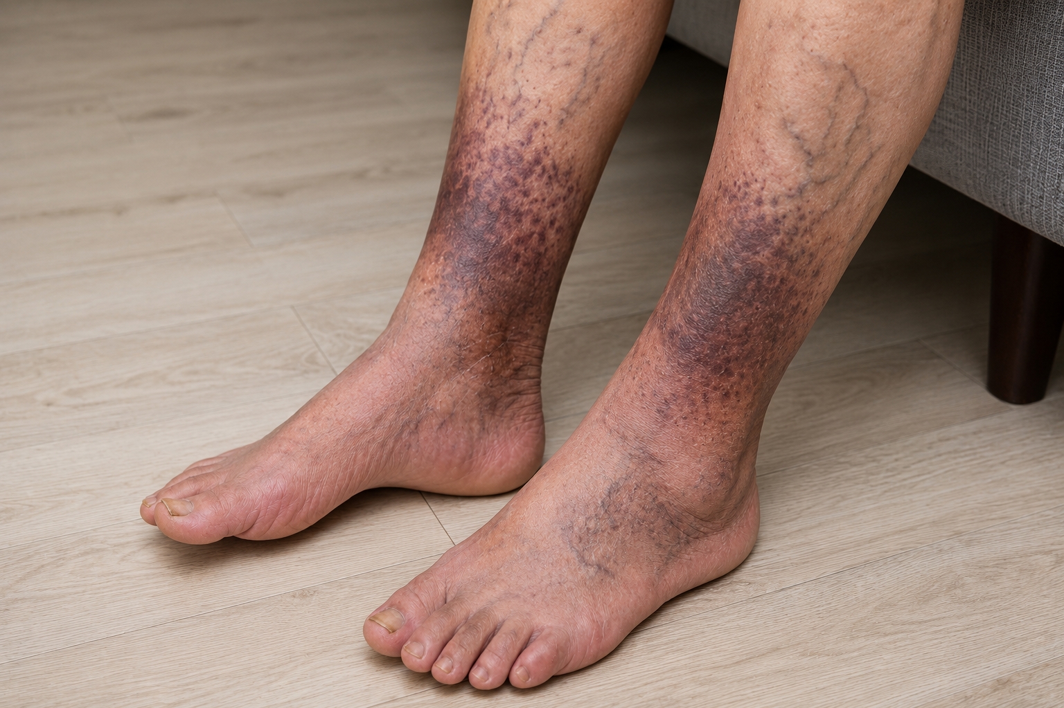
Red blood cells leak through these damaged capillaries into the surrounding tissue. Hemoglobin degrades into hemosiderin (iron deposits), producing the characteristic brown skin discoloration seen around venous ulcers.
Iron overload locks macrophages in an M1 (pro-inflammatory) state, driving tissue destruction rather than repair. Matrix metalloproteinases (MMPs) — enzymes normally involved in wound remodeling — become overexpressed and destroy the collagen and extracellular matrix needed for healing. Fibroblasts differentiate into contractile myofibroblasts that increase dermal tension.
This process establishes a self-perpetuating cycle in which chronic inflammation impairs healing and persistent venous hypertension continually reinjures the tissue. Without intervention to address the underlying venous pressure, wound closure cannot occur.
The CEAP classification
The CEAP classification system is used to grade the severity of chronic venous disease. Venous ulcers correspond to the most advanced stages: C5 indicates a healed venous ulcer, while C6 denotes an active (open) ulcer. However, pathological changes begin at earlier stages, including skin discoloration (C4a), lipodermatosclerosis (C4b), and corona phlebectatica (C4c), all of which serve as warning signs for potential ulceration if venous hypertension remains uncorrected.
Signs and symptoms
Characteristics of venous ulcers
Venous leg ulcers exhibit distinctive characteristics. They are typically located in the gaiter zone (ankle to mid-calf, usually on the medial side), are shallow with irregular borders, and have a wound bed containing pink or yellow granulation tissue, in contrast to the pale or black tissue of arterial ulcers. These ulcers often produce moderate to heavy exudate, and the surrounding skin may display hemosiderin staining, lipodermatosclerosis, or stasis dermatitis. Pruritus may occur, and pain levels vary; some ulcers are painful, particularly when infected, while others cause minimal discomfort.
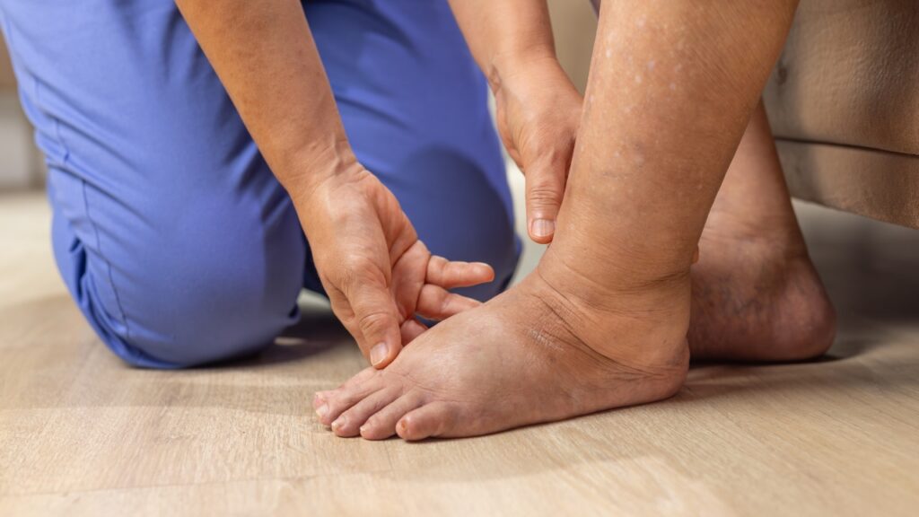
Warning signs that precede ulceration
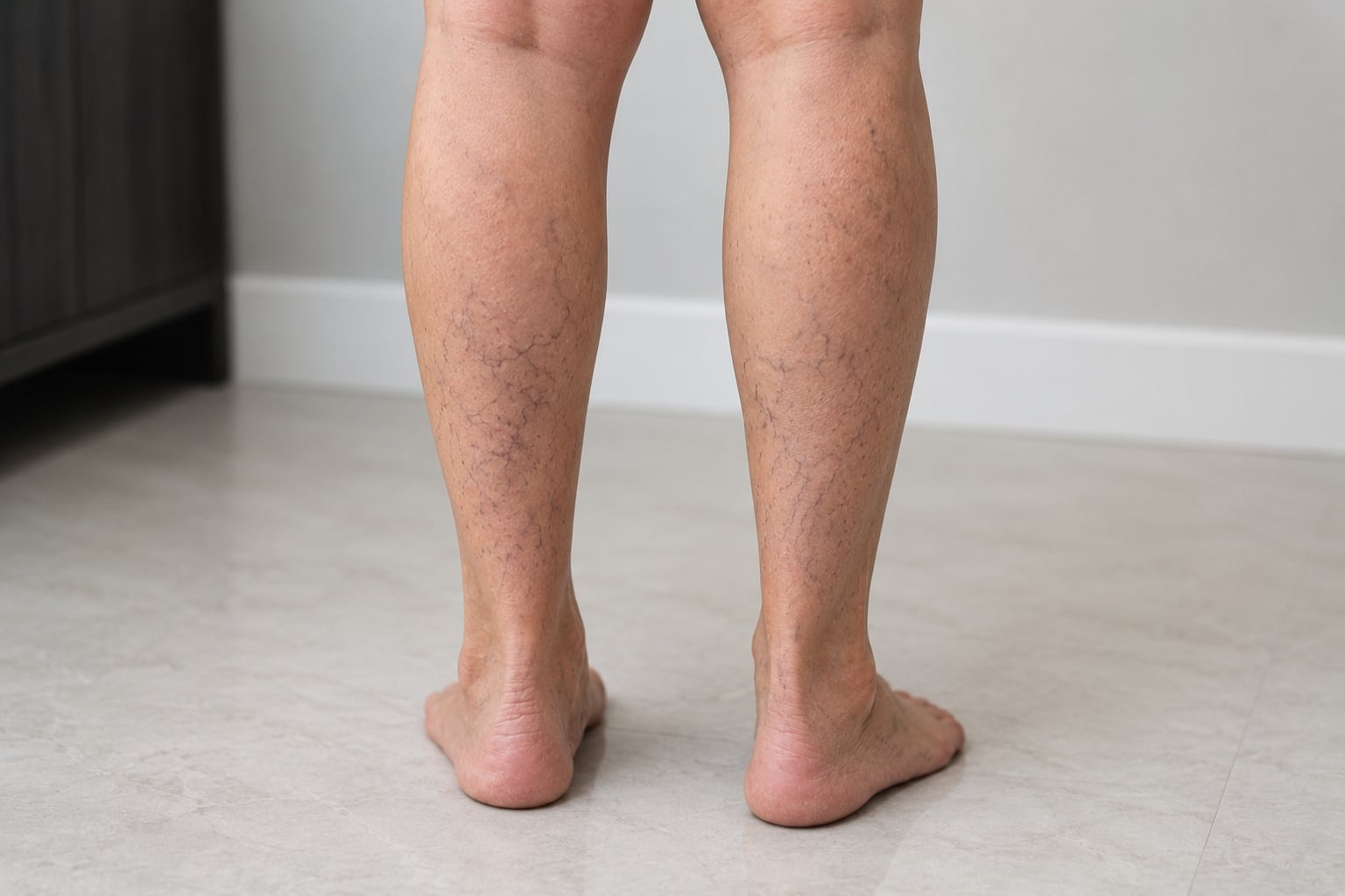
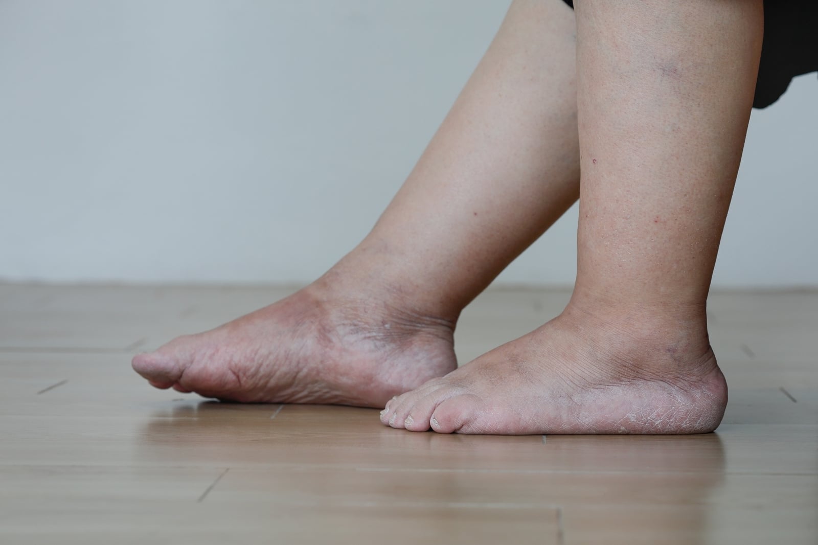
Venous ulcers are typically preceded by warning signs that may develop over months or years. These include persistent leg heaviness and fatigue, ankle swelling that worsens throughout the day, progressive skin darkening around the ankles, dry or flaky skin over the lower legs (stasis dermatitis), skin that becomes thick, hard, or woody (lipodermatosclerosis), and the presence of corona phlebectatica (fan-shaped spider veins around the ankle).
When to seek immediate medical attention
Any open wound on the leg that does not demonstrate improvement within two weeks should be evaluated by a wound care specialist or vein specialist. Urgent assessment is indicated if the wound exhibits signs of infection (such as increasing redness, warmth, swelling, purulent drainage, fever, or escalating pain), rapid expansion in size, surrounding skin that becomes hot and erythematous (suggestive of cellulitis), or if the wound occurs in individuals with diabetes, PAD, or immunocompromising conditions.
Diagnosis

Clinical assessment
Diagnosis commences with a comprehensive wound assessment, including evaluation of location, size, depth, wound bed characteristics, exudate, surrounding skin changes, and pain. The presence of hemosiderin staining, lipodermatosclerosis, and a medial gaiter-zone location strongly suggests a venous etiology.
Ankle-brachial index
The ABI is a critical assessment prior to initiating compression therapy. It compares ankle and brachial blood pressures to evaluate arterial sufficiency. An ABI below 0.8 indicates significant arterial disease and necessitates modified compression or arterial intervention before standard venous treatment. The EVRA trial excluded patients with an ABI below 0.8, underscoring the importance of this screening procedure.
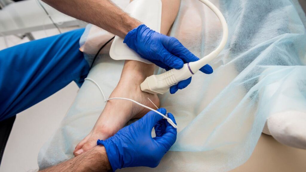
Duplex ultrasound
Duplex ultrasound is used to identify the specific veins contributing to venous hypertension. It delineates the location and severity of reflux within the superficial, deep, and perforator venous systems. This information is essential for treatment planning, as it determines which veins require ablation and whether deep venous disease, which may necessitate alternative management, is present.
Wound biopsy
A biopsy is indicated when an ulcer exhibits atypical features, fails to respond to standard treatment, or persists for an extended duration. This procedure is necessary to exclude malignancy (such as squamous cell carcinoma arising in chronic wounds, known as Marjolin ulcer), vasculitis, pyoderma gangrenosum, and other non-venous etiologies.
Treatment
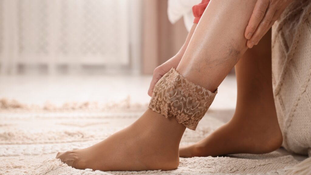
Compression therapy (Foundation of care)
Compression therapy is the cornerstone of venous ulcer management. A Cochrane Review of 22 trials demonstrated that compression increases ulcer healing rates compared with no compression, that multi-layered systems are more effective than single-layered systems, and that high compression is superior to low compression.
Compression therapy functions by counteracting venous hypertension, reducing edema, enhancing calf muscle pump efficacy, and promoting microcirculatory function. Available modalities include multi-layer compression bandaging (the traditional approach for active ulcers), compression stocking kits (two-layer systems designed for ease of application), and adjustable compression wraps (Velcro-based systems suitable for patients with limited dexterity).
Adequate arterial perfusion, defined as an ABI of 0.8 or higher, must be confirmed prior to the application of full compression therapy.
Early endovenous ablation (EVRA Evidence)
The EVRA trial, a landmark randomized controlled study involving 450 patients, fundamentally altered the management of venous ulcers. Patients assigned to early endovenous ablation of superficial venous reflux (within two weeks) in addition to compression therapy experienced significantly faster healing compared to those receiving compression alone with deferred ablation. The early intervention group also achieved longer ulcer-free intervals during the first year.
Long-term follow-up data (median 3.7 years) confirmed that early ablation reduced the overall incidence of ulcer recurrence and was highly cost-effective. The study authors recommended revising leg ulcer care pathways to incorporate early assessment and treatment of superficial venous reflux.
Ablation options include endovenous laser ablation (EVLA), radiofrequency ablation (RFA), ultrasound-guided foam sclerotherapy (UGFS), and cyanoacrylate glue closure. All were used in the EVRA trial.
Wound care
In addition to compression and venous intervention, the ulcer requires appropriate wound care. This encompasses regular wound cleansing, selection of suitable dressings (such as moisture-retentive dressings that maintain a moist environment while managing exudate), debridement of necrotic or non-viable tissue when indicated, and infection management (systemic antibiotics are reserved for cases of clinical infection, not for colonization alone).
Treating perforator and deep vein disease
A study involving VLU patients treated with radiofrequency ablation demonstrated that concomitant ultrasound-guided foam sclerotherapy of incompetent perforator veins at the time of saphenous ablation significantly improved healing rates. In this cohort, deep vein reflux was identified as the sole independent risk factor for ulcer recurrence (hazard ratio 3.72). These findings support a comprehensive treatment approach that addresses all sources of venous hypertension, rather than focusing solely on the saphenous trunk.
Skin grafting and advanced therapies
For ulcers unresponsive to compression and venous correction, advanced therapies may be considered. These include split-thickness skin grafting, bioengineered skin substitutes, negative-pressure wound therapy (wound VAC), and hyperbaric oxygen therapy, although evidence for the latter remains limited and its use is determined on a case-by-case basis.
Preventing recurrence

Venous ulcer recurrence represents a major challenge in long-term management. A longitudinal study of 250 patients reported recurrence rates of 22% at three months, 57% at one year, and 78% at three years. However, several modifiable factors can substantially reduce this risk.
Evidence-based preventive strategies
- Lifelong compression is the most important preventive measure. A 2024 Cochrane Review of eight studies involving 1,995 patients confirmed that compression reduces ulcer recurrence.
- Definitive venous intervention is also critical. A study of 1,324 ulcerated legs found that untreated superficial venous reflux was an independent risk factor for recurrence (hazard ratio 2.22). Addressing the underlying source of venous hypertension through ablation offers the most durable protection.
- Leg elevation for at least 30 minutes daily was independently associated with reduced recurrence (hazard ratio 0.33).
- Physical activity and higher self-efficacy (confidence in managing one’s condition) were also protective. Additional preventive strategies include weight management, skin care (such as moisturizing to prevent skin breakdown), and prompt treatment of new venous symptoms.
Frequently asked questions
With appropriate treatment (compression plus venous intervention), many ulcers heal within three to six months. Without treatment, healing can take years — and some ulcers never heal. The EVRA trial showed that early endovenous ablation significantly accelerated healing compared with compression alone.
Rarely. A chronic wound that has been present for many years can undergo malignant transformation (Marjolin ulcer). Any wound with atypical features, raised wound edges, or failure to respond to appropriate treatment should be biopsied.
Recurrence is a real risk — up to 78% within three years without sustained preventive care. However, combining definitive venous treatment (ablation), lifelong compression, regular exercise, and leg elevation substantially reduces this risk.
Standard high compression requires an ABI of 0.8 or higher. For patients with mild to moderate arterial disease (ABI 0.5–0.8), modified reduced-pressure compression may be used under specialist supervision. Compression is contraindicated in severe arterial disease (ABI below 0.5).
Both. Effective venous ulcer treatment requires wound care (compression, dressings, infection management) and vein treatment (addressing the underlying reflux). Many vein specialists coordinate with wound care teams or offer both services.
Bottom line
A non-healing open wound on the leg is among the most serious complications of untreated venous disease. Venous leg ulcers develop as a result of prolonged venous hypertension, which damages the skin and underlying tissues and establishes a chronic inflammatory environment that impedes healing.
The evidence is unequivocal: Compression therapy promotes venous ulcer healing, and early endovenous ablation of underlying venous reflux, as demonstrated by the EVRA trial in the New England Journal of Medicine, accelerates healing, increases ulcer-free intervals, and reduces recurrence. Early intervention is associated with improved outcomes.
If an open wound on the leg persists for more than two weeks, or if skin changes indicate progression of venous disease toward ulceration, prompt evaluation by a vein specialist is warranted.
- National Institutes of Health. 2026. Diabetic Foot Ulceration and Complications.
- National Institutes of Health. 2026. Peripheral Arterial Disease.
- PubMed Central. 2024. Prevention strategies for the recurrence of venous leg ulcers.
- PubMed Central. 2023. Prevalence and incidence of venous leg ulcers.
- PubMed. 2005. Chronic venous insufficiency.
- PubMed. 2024. Venous Insufficiency: Wound Management.
- PubMed. 2017. Pathophysiology of venous ulceration.
- PubMed. 2018. A Randomized Trial of Early Endovenous Ablation in Venous Ulceration.
- PubMed. 2012. Compression for venous leg ulcers.
- PubMed. 2014. Comparative systematic review and meta-analysis of compression modalities for the promotion of venous ulcer healing and reducing ulcer recurrence.
- PubMed. 2018. A Randomized Trial of Early Endovenous Ablation in Venous Ulceration.
- PubMed Central. 2020. Long-term Clinical and Cost-effectiveness of Early Endovenous Ablation in Venous Ulceration.
- PubMed. 2020. Factors that influence venous leg ulcer healing and recurrence rate after endovenous radiofrequency ablation.
- PubMed. 2015. Identifying risk factors and protective factors for venous leg ulcer recurrence using a theoretical approach.
- Cochrane. 2024. Compression therapy for preventing venous leg ulcers returning.
- PubMed. 2005. Risk factors for delayed healing and recurrence of chronic venous leg ulcers.
Editorial standards
All iThriveVeins content is medically reviewed by board-certified vein specialists and written following evidence-based guidelines. We source our information from peer-reviewed medical journals, clinical studies, and established medical organizations. Our editorial process ensures accuracy, objectivity, and relevance to patient needs.